While conventional examination, including Inspection, palpation (checking by touch) have their benefits, these are highly insignificant for the critical element – to identify dilated veins of normal function from dilated veins that carry blood in reverse direction
Duplex Scan is a combination of
- Doppler imaging: to see Function (direction of blood flow)
- 2-Dimentional ultrasound Imaging to show the anatomy
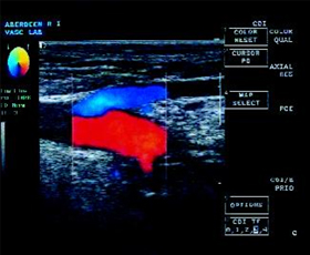
 Adjacent Image shows:
Red Colour – Forward flow in Common Femoral Vein (DEEP VEIN)
Blue Colour – Reverse flow in GSV (SUPERFICIAL VEIN)
This shows the vein at the SFJ (Sapheno Femoral Junction) is faulty, leading to Sapheno Femoral Incompetence.
This faulty venous pathway needs to be abolished/blocked to ensure venous blood flows only through competent pathways – reverse flow through incompetent pathways, aggravated during walking, lead to Ambulatory Venous Hypertension (AVH).
Duplex Scan is an integral part of evaluation, treatment and follow up evaluation
In this examination,
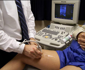
 Gel is applied to the skin of the leg. A transducer (a handheld device) is then used to scan the veins, by moving the probe over the skin above the vein.
- . The transducer sends high-frequency sound waves into the leg, and images of blood flow are obtained which show the direction of blood flow in the veins. This information is used to identify venous insufficiency (reflux).
- Ultrasound is then used to direct appropriate treatment of varicose veins.
Finally, evaluation with the same machine and same operator help in efficient treatment

